The above image demonstrates the collateral circulation associated with Moyamoya Disease.
This disease is very rare and is caused by a blockage of arteries in an area of the brain called the basal ganglia. In japanese moyamoya means, "puff of smoke" this refers to the way all the tiny vessels look that form around the blocked area. This disease is most common in children but is know to affect some adults. The associated symptoms of this disease are strokes or TIA's accompanied by muscular weakness, paralysis, or seizures. Adults with Moyamoya will most likely incurr an hemorrhagic strokes due to clots that form in the affected vessels. Those with Moyamoya will experience speech difficulties, disturbed consiousness, sensory and cognitive impairments, involuntary movements, and vision problems. Some researchers speculate that it is a genetic abnormality because it does tend to run in families. Revascularization surgery is usually the best form of treatment to open the blockages. Children usually respond better to theses surgeries than adults but for the most part neither should experience any strokes or TIA's. Without treatment Moyamoya is fatal becuase of intracerebral strokes.
radtpath23
Thursday, April 14, 2011
Wednesday, April 13, 2011
Facial Bones-blow out fracture
A blow out fracture occurs when there is trauma to the floor of the orbit. The trauma must exceed the tolerance of the bone. If this occurs with from a direct injury there is usually a naso-orbital fracture present as well. This is usually associated with blunt trauma and are often seen in motor vehicle accidents or an object striking the orbit. The symptoms of a orbital fracture are edema of the tissue lid, difficulty with vertical movement of the eye, or subcutaneous emphysema. A nose bleed could occur becuase of the connection with the maxilla and the nasal sinus. In severe cases of this fracture the orbit of the eye can actually drop into the maxillary sinus causing entrapment. This results in an inablitly to look superiorly or inferiorly. This can be corrected with surgery but if left unattended permnent stenosis of the muscle can occur. For diagnosis CT is the gold standard both axial and coronal scans should be obtained.
Nasal Polyps
The letter P demostrates the nasal polyp
- Nasal polypls occur when tissue becomes inflamed in the nasal mucosa or lining and form a sac-like growth. Polyps usally begin to occur by the ethmoid sinus and tend to grow in an open area. These polyps can eventually block the entire sinus or airway. Some of the common causes of these polyps are an aspirin sensitivity resulting in wheezing, asthma, chronic sinus infections, cycstic fibrosis, or hay fever (allergic rhinitis). The symptoms that tend to accompany polyps are a cold that will not go away, breathing only through the mouth, a nasal blockage, reduced or inability to smell, or runny nose. To diagnose polyps a CT is the gold standard, the polyps will show us as an opaque area on the scan. Treatment options are nasal steriod sprays but if discontinue use the symptoms will re occur, corticosteriod pills or liquid, antibiotics only if accompanied by a bacterial infection. In severe options individuals may need a surgical procedure called functional endoscopic sinus surgery. If surgery is indicated removal of the polyps will make it easier to breathe but the polyps will often return.
Monday, March 28, 2011
Brown's Syndrome
Brown's Syndrome is characterized by defects and errors in eye movement. This disease may be congential or secondary from inflammation. Malfunction of the superior orbital tendon causes these defects in eye movement, especially when adducting the eye upward. It is thought that about 35% of patients with Brown's Syndrome has a family member with Brown's Syndrome. It is thought then, that Brown's Syndrome could be a genetic trait. This syndrome is mostly found in women and then only in the right eye.
Some medical treatments for this syndrome are an antinflammatory such as ibuprofen. Another form of treatment is a steriod injection. However, surgical treatment is the best way to treat this syndrome. A tenotomy is performed and medical grade silicone 240 retinal band, this is the most effective form of treatment. Other surgical procedures include: superior oblique split tendon lengthening, tenotomy, and superior oblique recession. Facial reconstruction is also an option but it is not recommended for its low success rate. However, in some cases the best way to treat this syndrome is to do nothing at all!
Some medical treatments for this syndrome are an antinflammatory such as ibuprofen. Another form of treatment is a steriod injection. However, surgical treatment is the best way to treat this syndrome. A tenotomy is performed and medical grade silicone 240 retinal band, this is the most effective form of treatment. Other surgical procedures include: superior oblique split tendon lengthening, tenotomy, and superior oblique recession. Facial reconstruction is also an option but it is not recommended for its low success rate. However, in some cases the best way to treat this syndrome is to do nothing at all!
Monday, March 14, 2011
Temporal Bone Disease
What do you notice that is different about these images?? If you look closely at the arrows you will notice calcifications that should not be there. This is known as tympanosclerosis and by definition it is the formation of dense connective tissue in the middle ear, often resulting in hearing loss when the ossicles are involved. Tympanosclerosis is also known as myrinosclerosis or intratympanic tympanosclerosis. Myringosclerosis is classified by calcificaton only withing the tympanic membrane and and intratympanic tympanosclerosis is classified by calcification of any other structure in the middle ear, namely the ossicular chain, middle ear mucosa, or mastoid cavity. These diseases rarely result in any symptoms however, tympanoscerlosis can result in hearing loss or chalky white patches in the middle ear or temporal membrane. The exact cause of tympanosclersis is not completely understood but some probable factors are: long term otitis media, insertion of tympanostomy tubes, and atherosclerosis. Treatment for this disease can be hearing aids to treat the associated hearing loss or surgery. This involves removal of the affected areas and repair to the ossicular chain. Results are variable and can sometimes result in damage to the inner ear.
Tuesday, March 1, 2011
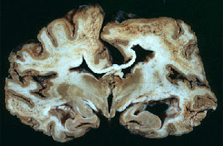
Creutzfeldt Jakob Disease
The above image is an actual brain with Creutzfeldt Jakob Diseae. Notice the large holes throughout the tissues!!
Creutzfeldt Jakob disease is a degenerative brain disorder that can lead to dementia and eventually death. This disease can resemble some dementia like brain disorder. However this disease will progress more rapidly than any type of dementia. In the 1990's this disease was on the rise in the United Kingdom. Individuals developed a form of this disease callled variant CJD. It was discovered that cattle had also contracted a form of this disease and the individuals most like contracted CJD from eating diseased meat. Fortunatly this disease is very rare with only one out of 1 million people being diagnosed each year. CJD has numerous symptoms and is therefore hard to diagnose; symptoms include: personality changes, anxiety, depression, memory loss, impaired thinking, blurred vision, insomnia, difficulty speaking and swallowing, and sudden jerky movements. As the disease progresses the mental symptoms may progress. CJD will usually cause the patient to go into a coma and heart failure, respitory failure, pneumonia, or other infections will likely be the actual cause of death. The estimated survival time is about 7 months.
CJD is caused by a varing group of both human and animal diseases known as transmissible spongiform encepholopathies. The disease name comes from the multiple holes that form in brain tissue during the progression of the disease. CJD is transmitted three ways. The majority of cases occur spontaneously with no clear reason for its development; this accounts for most of the diagnosed cases. Genetic mutation can also cause this disease as well as family history. This accounts for 5-10% of the cases. Lastly, individuals can be exposed by contamination of other human body tissue, or by eating contaminated meat from cattle with "mad cow disease".
Wednesday, February 23, 2011
Pituitary Microadenoma
A young man comes into the ER with vision changes. He has no other symptoms but is very frightened by these sudden changes!. After a stat CT head the Radiologist reveals that this young man has a pituitary microadenoma. A pituitary microadenoma is a tumor less than 10 mm in diameter. These adenomas are capable of secreting hormones or being clinically inactive. If this leasion is discovered while investigating other problems they are called incidentalomas. This tumor usually originates from a local mutation with loss of function of the genes controlling self proliferation. According to studies the United States has a 10% prevalence of pituitary leasions, and internationally an 11% prevelence. Microadenomas are not usually terminal but do put pressure onto to the optic chiasma; which can cause vision loss. These adenomas can occur at any age but do tend to occur in the aging population.
There are several different types of microademonas. If it is an incidentalomas there is usually no other symptoms and most times theses tumors are found in those who are seeking treatment for another condition such as headache. Prolactinomas may also be asymptomatic as long as the prolactin levels are not very elevated. In women these tumors can cause galactorrhea, amenorrhea, or infertility. In men this can cause hypogonadism, erectile dysfunction, and decreased libido. ACTH -secreting adenomas can cause Cushings Disease, Growth Hormone-Secreting adenomas cause acromegly, TSH-Secreting hormones are rare but will cause hypothyroidism.
Treatment for adenomas can range from medication to surgery to diet change. For prolactinomas, dopaminerginic drugs are given. For other tumors, especially those resulting in Cushnings and agromegaly, surgical removal is advised. Radiation therapy may also be indicated. Overall most microadenomas are too small to cause any problems and most are incidental findings but each case should be treated carefully and no assumptions should be made in regards to how an individual will react to treatment.
Subscribe to:
Comments (Atom)


